NEW YORK HOME X-RAY/US PROVIDES AN OUTREACH PORTABLE DIAGNOSTIC SERVICE FOR:
- Home Bound Patients
- The Elderly
- Nursing Homes
- Assisted Living Centers
- Occupational Screenings
- Facilities
- Other Settings by Arrangement
BY USING NEW YORK HOME X-RAY PORTABLE DIAGNOSTIC SERVICES YOU CAN:
Portable imaging keeps patients who are physically and/or psychologically impaired due to advanced age or physical limitations in their home vs transporting them to a hospital by ambulance. It also provides better patient care management through expedited radiology reports to their physicians, enabling doctors to provide immediate medical feedback.
X-RAYS ULTRASOUNDS EKGS TELEMETRY HOLTERS
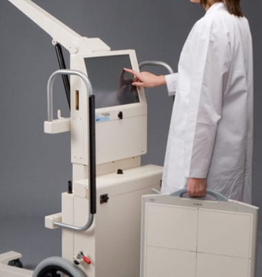


X-rays
Same Day Service
What is Plain radiography/X-rays?
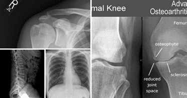
Radiography is the imaging of body structures using X-rays, which are a form of radiation similar to visible light, radio waves and microwaves. X-radiation is special because it has a very high energy level that allows the X-ray beam to penetrate through the body and create an image or picture.
The image is created due to the X-ray beam being absorbed differently by different structures or parts in the body. A dense structure like bone absorbs a high percentage of the X-ray beam (which appears light grey on the image), whilst low density structures like soft tissues absorb a small percentage (which appears dark grey on the image). The body has many different structures of varying densities and this difference creates a picture or image.
What are the risks of Plain radiography/X-rays?
Generally, the benefit of the X-ray procedure is far more important than the small estimated risk. At the radiation dose levels that are used in diagnostic radiography there is little or no evidence of health effects.
The two major risks to health that occur as a result of exposure to medical ionizing radiation (which is the kind of radiation in X-rays) are:
- Cancer occurring many years after the radiation exposure
- Health problems in the children born to people exposed to radiation because of damage to the reproductive cells in the body.
To put this into perspective, a patient would need to have approximately 38 chest X-rays to receive an amount of radiation similar to that of normal background radiation that everyone receives for one year from the environment.
Who does Plain radiography/X-rays?
A radiographer or medical imaging technologist is a health licensed board certified professional who performs diagnostic radiography. A radiologist is a specialist medical doctor who reviews and interprets the images and provides a written report of the test to your referring doctor.
How do I prepare for Plain radiography/X-rays?
For a plain X-ray are no specific preparation instructions.
Please inform the radiographer who is performing the X-ray if there is any chance you may be pregnant. Safety of the patient and unborn child is the number one priority so a different approach or test may be needed.
- Be prepared to remove certain items like watches, necklaces and certain types of clothing that contain metal objects such as zips, as these items may interfere with the quality of the image.
What happens during Plain radiography/X-rays?
Depending on the part of your body being examined you may be asked to stand, sit or lie down while the X-ray is taken. The number of X-rays taken and the speed of the test will also depend on this. It is important that you stay completely still when the radiographer instructs you to, as any movement may create a blurred image.
After the X-rays have been performed, the radiographer has to process each X-ray and check the results for quality. This can sometimes take several minutes. Sometimes there will be a need for additional images to be taken to obtain more information to help the radiologist (a specialist doctor) make a diagnosis. There is no need for concern if this happens as it is quite common.
The radiographer will instruct you when the procedure is finished. The radiologist then carefully assesses the images, makes a diagnosis and produces a written report on the findings, which is sent to your referring doctor.
The entire process is straightforward and you will not feel anything strange or feel any different during the examination. You are welcome to ask questions at any stage.
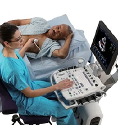
What is an Ultrasound?
Ultrasound is safe and painless and produces pictures of the inside of the body using sound waves. Ultrasound imaging, also called ultrasound scanning or sonography, involves the use of a small transducer (probe) and ultrasound gel placed directly on the skin. High-frequency sound waves are transmitted from the probe through the gel into the body. The transducer collects the sounds that bounce back and a computer then uses those sound waves to create an image. Ultrasound examinations do not use ionizing radiation (as used in x-rays), thus there is no radiation exposure to the patient. Because ultrasound images are captured in real-time, they can show the structure and movement of the body's internal organs, as well as blood flowing through blood vessels.
Ultrasound imaging is a noninvasive medical test that helps physicians diagnose and treat medical conditions.
Conventional ultrasound displays the images in thin, flat sections of the body. Advancements in ultrasound technology include three-dimensional (3-D) ultrasound that formats the sound wave data into 3-D images.
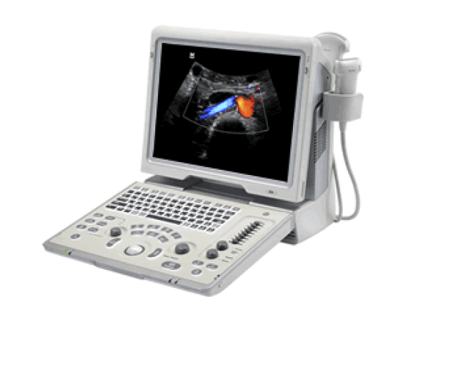
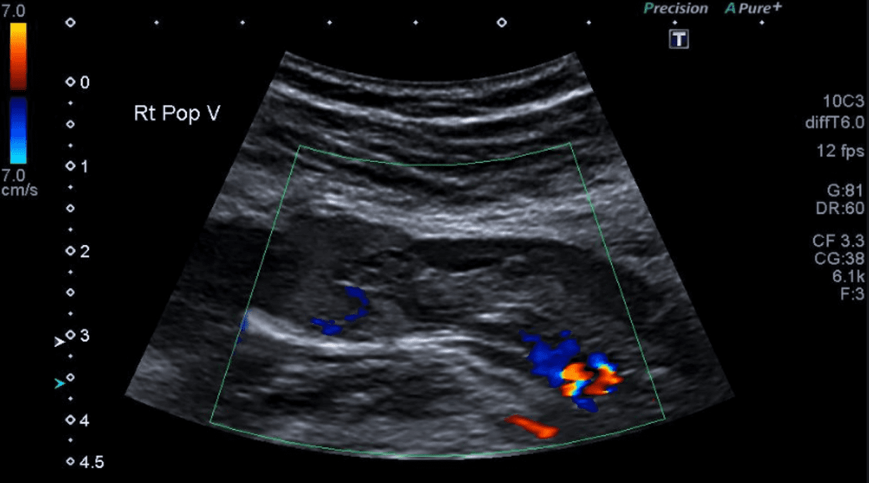
A Doppler ultrasound study may be part of an ultrasound examination.
Doppler ultrasound, also called color Doppler ultrasonography, is a special ultrasound technique that allows the physician to see and evaluate blood flow through arteries and veins in the abdomen, arms, legs, neck and/or brain (in infants and children) or within various body organs such as the liver or kidneys.
There are three types of Doppler ultrasound:
- Color Doppler uses a computer to convert Doppler measurements into an array of colors to show the speed and direction of blood flow through a blood vessel.
- Power Doppler is a newer technique that is more sensitive than color Doppler and capable of providing greater detail of blood flow, especially when blood flow is little or minimal. Power Doppler, however, does not help the radiologist determine the direction of blood flow, which may be important in some situations.
- Spectral Doppler displays blood flow measurements graphically, in terms of the distance traveled per unit of time, rather than as a color picture. It can also convert blood flow information into a distinctive sound that can be heard with every heartbeat.
What are some common uses of the procedure?
Ultrasound examinations can help to diagnose a variety of conditions and to assess organ damage following illness.
Ultrasound is used to help physicians evaluate symptoms such as:
- pain
- swelling
- infection
Ultrasound is a useful way of examining many of the body's internal organs, including but not limited to the:
- heart and blood vessels, including the abdominal aorta and its major branches
- liver
- gallbladder
- spleen
- pancreas
- kidneys
- bladder
- uterus, ovaries, and unborn child (fetus) in pregnant patients
- eyes
- thyroid and parathyroid glands
- scrotum (testicles)
- brain in infants
- hips in infants
- spine in infants
Doppler ultrasound images can help the physician to see and evaluate:
- blockages to blood flow (such as clots)
- narrowing of vessels
- tumors and congenital vascular malformations
- reduced or absent blood flow to various organs
- greater than normal blood flow to different areas, which is sometimes seen in infections
With knowledge about the speed and volume of blood flow gained from a Doppler ultrasound image, the physician can often determine whether a patient is a good candidate for a procedure like angioplasty.